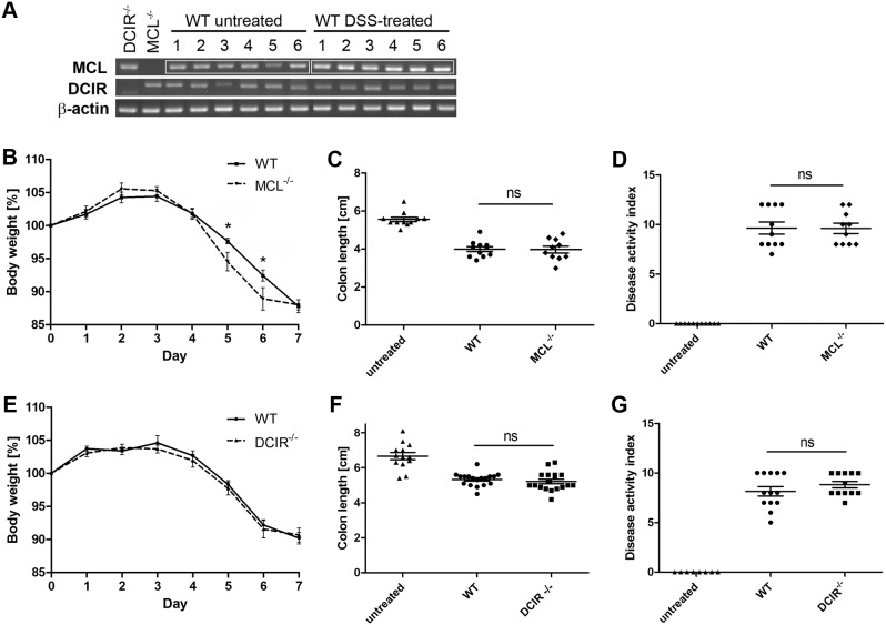
Figure 2. Clinical symptoms of DSS-induced colitis in MCL−/− and DCIR−/− mice.
Colitis was induced in wild-type, MCL−/− and DCIR−/− mice by adding 3% DSS into the drinking water for seven consecutive days. (A) MCL and DCIR mRNA levels were analyzed by semi-quantitative RT-PCR in colon of untreated and DSS-treated wild-type mice (6 mice per group). As a control, RNA was also isolated from colon of untreated MCL−/− or DCIR−/− mice. β-actin expression levels were determined in all samples to ensure the same quantity and quality of cDNA. MCL mRNA expression in colon of DSS-treated mice was increased compared to the colon of untreated mice while DCIR mRNA levels did not differ between untreated and DSS-treated mice. Disease severity was determined by the daily body weight loss for MCL−/− mice (n = 11) (B), and for DCIR−/− mice (n = 18) (E), the colon length for MCL−/− mice (C), and for DCIR−/− mice (F), and a disease activity index (DAI) based on blood occurrence, stool consistency and body weight loss for MCL−/− mice (n = 11 for wild-type and n = 10 for MCL−/− mice) (D), and for DCIR−/− mice (n = 13 for wild-type and n = 12 for DCIR−/− mice) (G). Data shown are the combined results of two (MCL) or three (DCIR) independent experiments. On day six, one MCL−/− mouse had to be euthanized due to the defined humane endpoints. MCL−/− mice displayed an increased body weight loss compared to wild-type mice, with a significant difference on day 5 and 6. The colon length and DAI did not vary between MCL−/− and wild-type mice. No significant difference in body weight, colon length, and DAI between DCIR−/− and wild-type mice was observed. Data are expressed as mean + SEM. The p-values were determined with Mann-Whitney’s U test (*p<0.05). Significance is indicated by asterisks (*), ns = no significance.

