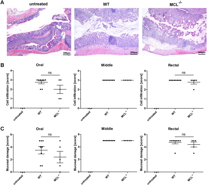
Figure 3. Histological analysis of colon sections from wild-type and MCL−/− mice.
Paraffin sections of the colon from untreated or 3% DSS-treated wild-type and MCL−/− mice were prepared at day seven and were stained with hematoxylin and eosin (H&E) for histological evaluation in a blinded manner. Each colon was divided into three segments of identical length (oral, middle, rectal) which were separately analyzed. (A) Representative images of paraffin-embedded sections of the rectal part of the colon are shown (40x magnification). The degree of leukocyte infiltration (B) and mucosal erosion/ulceration (C) was graded from none (score 0) to mild (score 1), moderate (score 3), or severe (score 4). Data are expressed as mean + SEM (n = 8 for wild-type and n = 5 for MCL−/− mice). The p-values were determined using Mann-Whitney’s U test. Significance is indicated by asterisks (*), ns = no significance.

