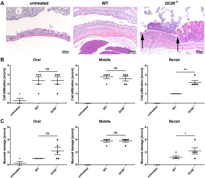
Figure 4. Histological analysis of colon sections from wild-type and DCIR−/− mice.
Paraffin sections of the colon from untreated or 3% DSS-treated wild-type and DCIR−/− mice were prepared at day seven and were stained with hematoxylin and eosin (H&E) for histological evaluation in a blinded manner. (A) Representative images of paraffin-embedded sections of the rectal part of the colon are shown (40x magnification). Arrows indicate a severe ulcer in the colon from DCIR−/− mice. Each colon was divided into three segments of identical length (oral, middle, rectal) which were separately analyzed. The degree of leukocyte infiltration (B) and mucosal erosion/ulceration (C) was graded from none (score 0) to mild (score 1), moderate (score 3), or severe (score 4). The scores for both, cell infiltration as well as mucosal ulceration in the rectal part of the colon from DCIR−/− mice were significantly increased compared to wild-type mice. Data are expressed as mean + SEM (n = 5). The p-values were determined using Mann-Whitney’s U test (*p<0.05, **p<0.01). Significance is indicated by asterisks (*), ns = no significance.

