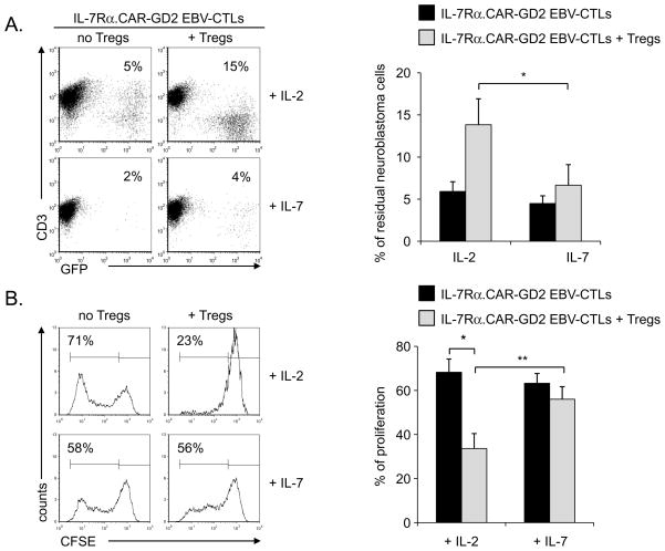
Figure 4. IL-7, unlike IL-2, supports in vitro the proliferation and function of IL-7Rα.CAR-GD2+ EBV-CTLs in the presence of Tregs.
Panel A. IL-7Rα.CAR-GD2+ EBV-CTLs were co-cultured with CHLA-255 GFP-tagged cells (ratio 1:2) in the presence of IL-2 or IL-7, with or without Tregs. The percentage of residual tumor cells was measured by flow cytometry on day 7 of culture. The plots on the left show a representative experiment, while the graph on the right summarizes mean ± SD of 5 independent experiments. Panel B. IL-7Rα.CAR-GD2+ EBV-CTLs were labeled with CFSE and activated with autologous LCLs in the presence of IL-2 (upper plots) or IL-7 (lower plots) with or without Tregs. CFSE dilution was measured at day 7 of culture by flow cytometry. The plots on the left show a representative experiment, while the graph represents mean ± SD of 5 independent experiments. * p<0.01; ** p=0.005

