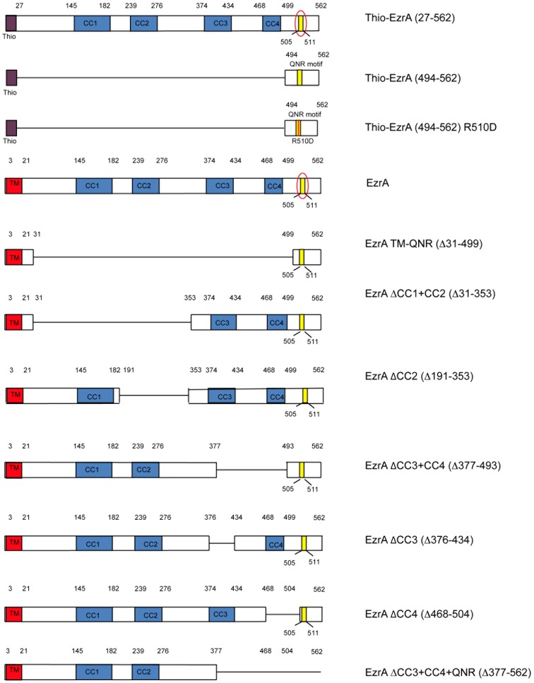
Figure 1. Schematic of EzrA deletion mutants employed in this study.
The predicted coiled-coil structure of EzrA is drawn to scale. Numbers refer to amino acid positions. Coiled-coil regions are shaded blue. The QNR motif is shaded yellow and circled in the full-length protein. Lines indicate deleted regions. The QNR domain construct missing all four coiled-coils is just under the full-length protein. All constructs used for in vivo analysis were fused in-frame to a C-terminal GFP moiety (not shown) to simplify analysis. All constructs used for in vitro analysis were fused in-frame to a N-terminal Thio tag to enhance solubility and a N-terminal 6x-His tag for purification purposes (not shown).

