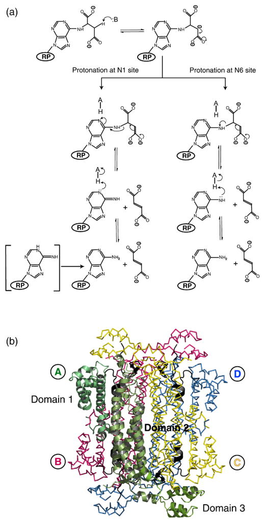
Figure 1.
(a) Proposed reaction scheme for ADL. The ribose and phosphate groups of ADS are represented by RP. (b) The quaternary structure of E. coli ADL. Monomer A is shown in cartoon representation with domains 1–3 (residues 1–121, 122–381, and 382–456, respectively) indicated. The regions of highly conserved amino acid residues, which form the active sites of the protein, are colored black (C1, residues 117–130; C2, residues 168–178; C3, residues 294–307). Monomers A, B, C and D are labeled and are colored green, pink, yellow, and blue, respectively. PyMol was used for Figure preparation [http://pymol.sourceforge.net/].

