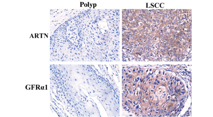
Figure 1.
Expression of ARTN and GFRα1 in LSCC and polyp tissues specimens. Immunohistochemical analysis of ARTN and GFRα1 protein in LSCC and polyp. Low expression of ARTN and GFRα1 in polyp (left panels). High expression of ARTN and GFRα1 in LSCC (right panels). All images are counterstained with hematoxylin. Photomicrographs were captured at ×200 magnification. LSCC, laryngeal squamous cell carcinoma; ARTN, artemin; GFRα1, GDNF receptor α1.

