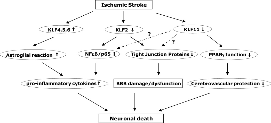
Figure 1.
Schematic representation of stroke-associated KLFs and signaling cascades. KLF4 is induced in reactive astrocytes in rat hippocampus after global cerebral ischemia, and may transcriptionally regulate astroglial reaction and cerebral inflammation following ischemic injury (Park et al., 2013). KLF2 level is decreased in blood outgrowth endothelial cells (BOECs) derived from stroke children with sickle cell anemia (SCA) than in non-SCA BOECs, thus changes the ratio between proinflammatory NF-κB/p65 to anti-inflammatory factor KLF2 and leads to a proinflammatory phenotype within the cerebral vasculature (Enenstein et al., 2010). In mouse brain vasculature, ischemic stimuli induce KLF2 (Shi et al., 2013) or KLF11 dysfunction (Yin et al., 2013), featuring attenuated KLF2 transactivation of tight junction protein occludin or reduced KLF11-mediated PPARγ vascular function, thereby resulting in a series of cerebral vascular pathologies, such as BBB damage/dysfunction, disturbed cerebral vascular protection, and final neuronal cell death.

