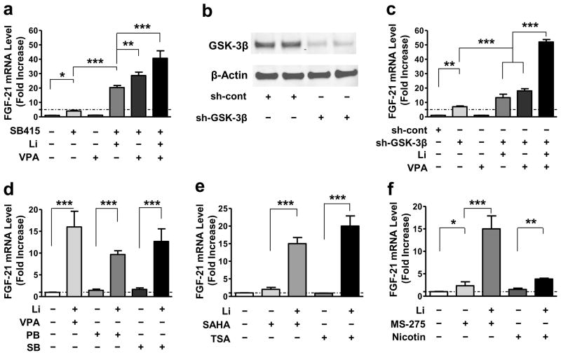
Figure 2. FGF-21 mRNA levels were increased by GSK-3 inhibition, and boosted by a combination of GSK-3 and HDAC inhibition.
(a) Starting at DIV-6, CGCs were treated with 10 μM SB415286 (SB415), 0.8 mM VPA, or a combination of 10 μM SB415 with 3 mM Li, 0.8 mM VPA, or with both 3 mM Li and 0.8 mM VPA for 48 hours, then harvested to detect FGF-21 mRNA level by q-PCR. (b) CGCs were transduced with sh-GSK-3β (#614) or sh-cont construct at the time of plating. At DIV-6, cells were harvested for Western blotting of total GSK-3β and β-actin protein levels. (c) CGCs transduced with sh-cont or sh-GSK-3β were treated with 3 mM Li, 0.8 mM VPA, or their combination, starting at DIV-6. Two days later, cells were harvested and q-PCR was performed to detect FGF-21 mRNA levels. (d) CGCs at DIV-6 were treated with 3 mM Li in the absence or presence of 0.8 mM VPA, 1 mM PB, or 1 mM SB. (e) CGCs at DIV-6 were treated with 3 mM Li in the absence or presence of 10 μM vorinostat (SAHA), 100 nM trichostatin A (TSA). (f) CGCs at DIV-6 were treated with 3 mM Li in the absence or presence of 5 μM MS-275, or 10 mM nicotinamide (Nicotin). The effects of these HDAC inhibitors alone were also assessed. Quantified data are means ± SEM and analyzed by one-way ANOVA, n=3; *P<0.05; **P<0.01; ***P<0.001.

