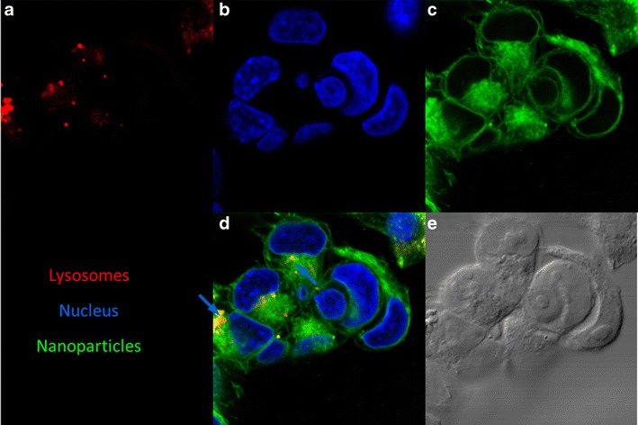
Fig. 5.

Intracellular distribution of PLGA nanoparticles. Confocal microscopic images of MA148 cells incubated with coumarin-6 labeled nanoparticles (green). Lysosomes were stained with LysoTracker red® (red) and nucleus with DAPI (blue). Representative images showing the location of a lysosomes, b nucleus, c nanoparticles, and d their colocalization. The merged image shows nanoparticles in lysosomes (blue arrow) and in the cytoplasm. e DIC image of the cells are also shown
