Fig. 6.

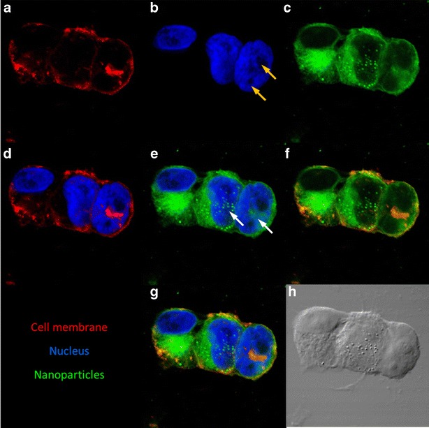
PLGA nanoparticles can enter the nucleus of dividing cells. Confocal microscopic images of MA148 cells treated with coumarin-6 labeled nanoparticles (green). Cell membrane was stained with Texas red®-X conjugated wheat germ agglutinin (red) and nucleus was stained with DAPI (blue). Representative images showing the individual staining of a cell membrane, b nucleus, c nanoparticles, merged image of d cell membrane and nucleus, e nanoparticles and nucleus, f nanoparticles and cell membrane, and g their colocalization. Presence of two nucleoli (yellow arrows) suggests cell division. White arrows indicate the presence of nanoparticles in the nuclear compartment of the cells. Absence of nanoparticles in nondividing cells (left most cell) and their presence in dividing cells indicate the possibility of nanoparticle entry into the nucleus of the rapidly dividing cells h Differential interference contrast (DIC) image of the cells
