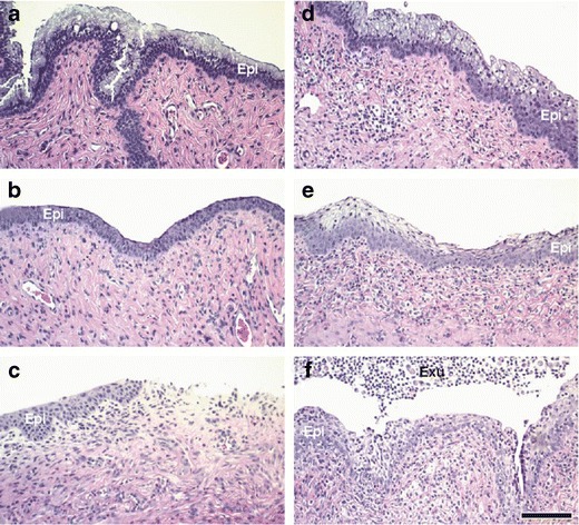
Fig. 8.

Progesterone-conditioned CD-1 mice were treated intravaginally with a carboxymethylcellulose (CMC) vehicle, b 8.0% N-9, c 2.0% BZK, d TA 1/SR-2P (1% Pluronic® F-127/ 1% Noveon® AA-1), e TA 2 (0.5% Pluronic® F-127/ 7.5% Noveon® AA-1), and f TA 3 (0.5% Pluronic® F-127/ 4.5% Noveon® AA-1) once daily for 12 days. Vaginal tissues were collected 1 day after last dose application, fixed in neutral-buffered formalin, and stained with H&E. After treatment with known vaginal irritants, epithelial thinning (b, c), erosion (c), leukocyte infiltration (b, c), and loss of mucification (b, c) were observed when compared to vehicle-treated animals (a). TA 2 and TA 3 application resulted in leukocyte infiltration and few minimal changes (e, f). In contrast, only minimal changes were observed after SR-2P/TA 1 treatment (d). Epi epithelium, Exu exudate; bar 100 μm
