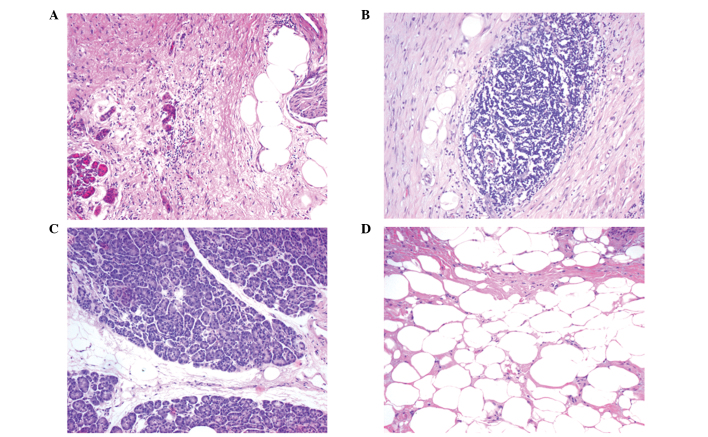
Figure 1.
Original grading examples of the histological analysis of the resection edges (hematoxylin and eosin stain; magnification, ×100). (A) Lipomatous atrophy grade 1; (B) lipomatous atrophy grade 3; (C) chronic inflammatory infiltration grade 1; and (D) chronic inflammatory infiltration grade 3.

