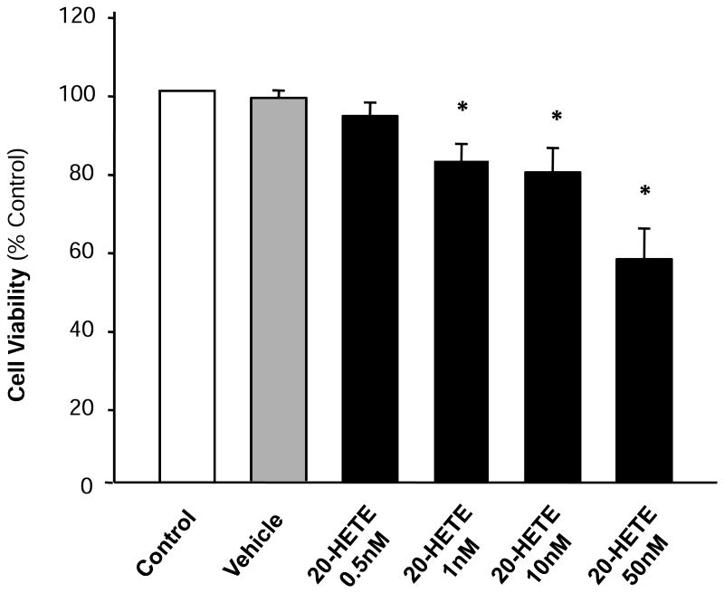
Figure 1. Effect of 20-HETE on the survival rate of cultured neonatal rat ventricular myocytes.
Serum deprivated cardiomyocytes were treated with control, vehicle, or 20-HETE (0.5 nM, 1 nM, 10 nM, 50 nM) for 24h. Cell viability was determined using the MTT assay as detailed in the Methods. Values are expressed for each time point as percentage of control. Data are mean±SE obtained from three experiments using independent batches of cells in each group. *P<0.05 compared with control.

