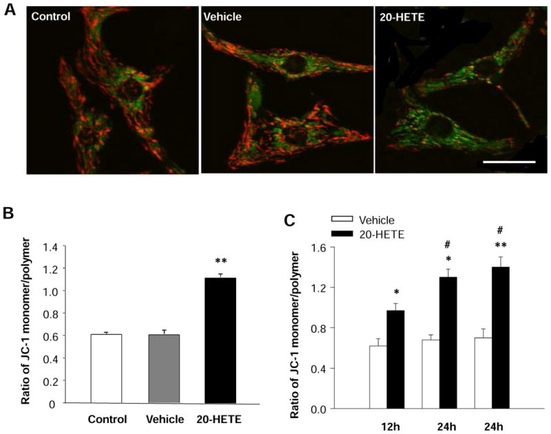
Figure 3. Effects of 20-HETE on the mitochondrial membrane potential (ΔΨm) of cultured neonatal rat ventricular myocytes.

Red fluorescence represents the mitochondrial aggregate form of JC-1, indicating intact mitochondrial membrane potential. Green fluorescence represents the monomeric form of JC-1, indicating dissipation of ΔΨm. A. JC-1 staining of control cardiomyocytes, exhibiting a heterogeneous distribution of mitochondria with low density of green fluorescence and high density of red fluorescence. The green fluorescence intensity was increased and the red fluorescence intensity was decreased by treatment with 20-HETE. Scale bar =20 μm. B. Quantitative analysis of JC-1 fluorescence in cardiomyocyte treated with vehicle control and 20-HETE (10 nM) for 24 h. Treatment of cell with 20-HETE significantly increased the ratio of green to red fluorescence, suggesting a depolarization of ΔΨm. Data are mean±SE obtained from 16 observation fields in three independent experiments in each group. **P<0.01 significant difference compared with control. C. Time-course of the effect of 20-HETE on the ratio of JC-1 monomer/polymer in cardiomyocytes. Data are mean±SE obtained in three independent experiments in each group. *P<0.05 significant difference compared with control. **P<0.01 significant difference compared with control. #P<0.05 compared with treatment with 20-HETE for 12 h.
