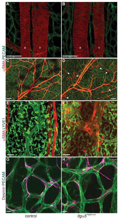
Figure 2. Normal blood vessel morphology in Itga5Pdgfrb-cre mice.
(A–H) Whole-mount immunofluorescence stainings of control and mutant embryonic skin at E17.5. (A, B) No obvious defects seen in vSMC (red) attachment or morphology around arteries (a) and veins (v) in Itga5Pdgfrb-cre mice. (C, D) Low magnification images showing the extent of vSMC coverage throughout the vasculature in a control (C) and Itg α5Pdgfrb-cre embryo (D). Notice the ectopic vSMC coverage around lymphatic capillaries (white arrows) (D), which are absent in control embryos (C). Higher magnification image of a lymphatic capillary (green) in control skin (E) and ectopic αSMA coverage around a lymphatic capillary in an Itg α5Pdgfrb-cre embryo (F). Desmin-positive pericytes apposed to microvessels in control (G) and Itg α5Pdgfrb-cre mutants (H). Scale bars: 50 μm (A, B,), 200 μm (C, D), 50 μm (E, F), 10 μm (G, H).

