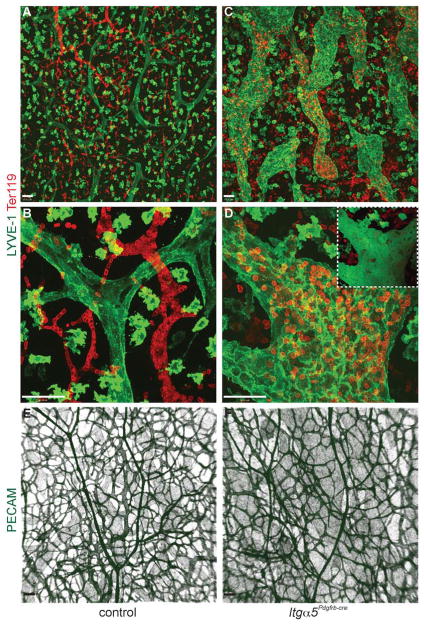
Figure 3. Itg α5Pdgfrb-cre mice display hyperplastic, blood-filled lymphatic vessels.
(A–D) Confocal images showing the lymphatic capillaries (LYVE-1, green; note that LYVE-1 is also expressed on macrophages) and erythrocytes (Ter119, red) in E15.5 control and Itg α5Pdgfrb-cre skin. In contrast to control mice (A and B), lymphatic vessels in Itg α5Pdgfrb-cre mutants are hyperplastic, tortuous and filled with red blood cells (C and D). Inset shows 3D rendering of image in (D) confirming presence of blood within the lymphatic vessel. Blood vessels in Itg α5Pdgfrb-cre mice (strong PECAM stain, green) however remain unaffected by the deletion of integrin-α5 (E, F). Note lymphatic vessels (weak PECAM stain) are also visible in both E and F. Scale bars: 50 μm.

