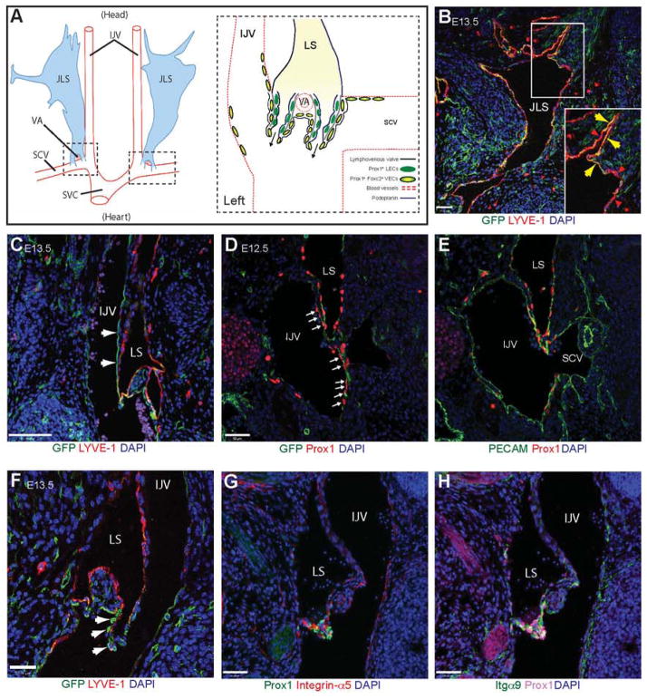
Figure 4. Lymphatic expression of Pdgfrb-Cre.
(A) Schematic representation of the position (modified from Srinivasan and Oliver (2011)) and morphology of the jugular lymphatic sacs (JLS) and the lymphovenous valves in E13.5 mouse embryos. The head is orientated to the top and the heart towards the bottom of the figure. The JLS runs from the neck posteriorly to the level of the thymus, where it is split into two portions by the vertebral artery (VA). The lymphovenous valves (dashed boxes) form at the end of these two lymph sacs (LS) through the fusion of the posterior region of the LSs with the internal jugular (IJV) and subclavian (SCV) veins where they merge and drain into the superior vena cava (SVC). Specialised Prox1+ Foxc2+ podoplanin− venous endothelial cells (VECs) are found in these regions. The lymphovenous valve leaflets consist of two layers of Prox1+ cells. The inner layer is composed of Prox1+ Foxc2− podoplanin− lymphatic endothelial cells (LEC) from the LSs, while the outer layer is derived from the Prox1+ Foxc2+ podoplanin− VECs. (B) Immunofluorescence staining on a coronal section through the JLS of a Pdgfrbmtmg mouse at E13.5 (Pdgfrβ+ cells will be GFP+). Note that the JLS appears to contain both GFP+ (yellow, arrows) and GFP− (red, arrowheads) lymphatic endothelial cells (B, enlarged in inset). (C) GFP+ cells are also present in a population of cells in the IJV and SCV (arrows), adjacent to the JLS where the LS merge with venous circulation to form the lymphovenous valves. (D, E) Sequential transverse sections stained with antibodies against the transcription factor Prox1 and (D) GFP and (E) PECAM showing Pdgfrb-Cre expression in the LS and in a specialised population of venous endothelial cells within the IJV and SCV (arrows) in an E12.5 Pdgfrbmtmg embryo. Note the lack of Pdgfrb-Cre expression in Prox1-negative venous endothelium. (F) Pdgfrb+ cells (arrows) in the outer layer of the lymphovenous valve. (G, H) Immunofluorescence staining on a cryosection through the lymphovenous valve showing high expression of (G) integrin-α5 and (H) integrin-α9 within the Prox1+ valve leaflets. Scale bars: 50 μm.

