
Figure 1.
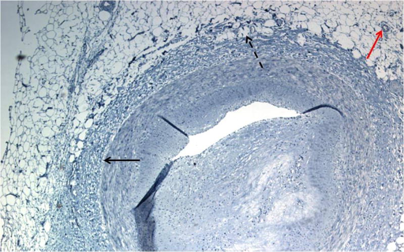
Histological appearance of human perivascular adipocytes surrounding an atherosclerotic coronary artery. Artery was fixed in formalin, sectioned, and counterstained with hematoxylin. Red arrow points to a blood vessel, solid black arrow denotes location of external elastic lamina, and dashed arrow points to PV adipocytes invading the lamina adventitia lacking a separating fascia. Note that these perivascular adipocytes exhibit an appearance more consistent with unilocular white adipocytes rather than multilocular brown adipocytes.