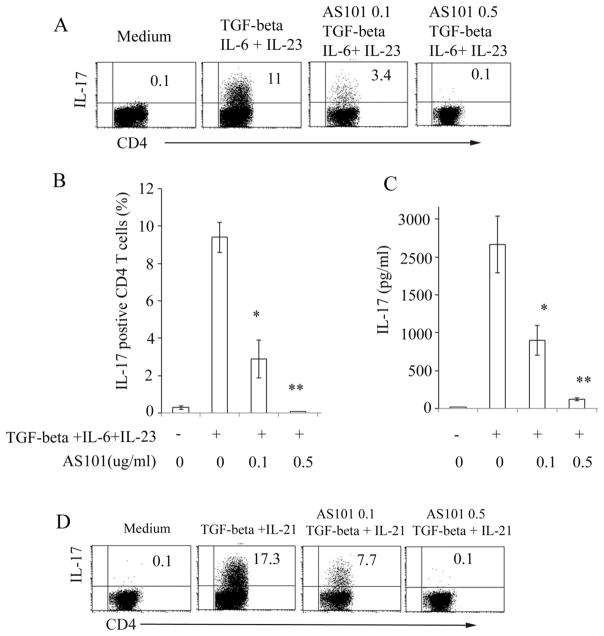
Figure 1. AS101 inhibits production of IL-17 by CD4+ T cells.
Isolated CD4+ T cells from 8 to 12 week old naïve C57BL/6 mice were cultured in 24 well plates coated with anti-CD3 (10μg/ml) under Th17 polarizing conditions (TGF-β 2 ng/ml +IL-6 20ng/ml + IL-23 20ng/ml) in the presence or absence of AS101 (0.1 to 0.5μg/ml). Anti-IFN-γ (10μg/ml), anti-IL-4 (5μg/ml), and anti-CD28 (3μg/ml) were added to the cultures. Intracellular cytokine staining was performed following 5 hrs restimulation with PMA/ionomycin/brefeldin A. FACSCalibur was used for the data collection (A). Summary of three representative experiments (Mean ± SEM) is shown (B). C. CD4+ T cell culture supernatants were harvested after 72 hrs culture and analyzed by IL-17 ELISA kits (eBiosciences). Summary of 3 individual experiments is shown (Mean ± SEM). D. Isolated CD4+ T cells were stimulated with different Th17 polarizing cytokines (TGF-β (2 ng/ml), IL-21 (50 ng/ml) in the presence or absence of AS101(0.1, 0.5μg/ml) as in Fig. 1A. *, p<0.05; **, p<0.01.

