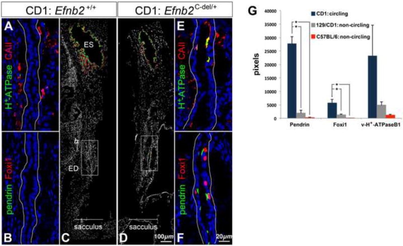
Figure 8. Mis-localization of Foxi1 and ion transport proteins in Efnb2c-del/+ fetuses of the circling CD1 strain.

(A-C) Saggital sections of an E19 CD1 wild-type head, double-immunolabeled for v-H+-ATPase and Carbonic anhydrase II (A) or pendrin and Foxi1 (B,C). (B) corresponds to the box in (C). (D-F) Saggital sections of an E19 CD1 Efnb2c-del/+ head, double immunolabeled for pendrin and Foxi1 (D,F) or v-H+-ATPase and Carbonic anhydrase II (E). (F) corresponds to the box in (D). Ductal epithelia (A,B,E,F) are outlined in white. (G) Sums of all positive pixels (means and standard deviations; n = 4 per genotype) for epitopes of interest from the proximal half of the ED in heterozygote fetuses at E19. Asterisks show significant differences (P < 0.05) by Tukey's post-hoc multiple comparison tests.
