Figure 6.
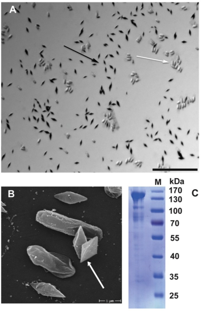
B. thuringiensis 4A4 produces crystal proteins. (A) Light microscopy image of Coomassie-stained spore-crystal mixture of B. thuringiensis 4A4. Spores are unstained (white arrow), black structures are crystal proteins (black arrow). Scale bar is 20 μm; (B) Scanning electron microscopy image of spore-crystal mixture of B. thuringiensis 4A4. Bi-pyramidal crystals are shown (pointed with white arrow); (C) SDS-PAGE profile of B. thuringiensis 4A4 crystal proteins. Dominant protein of around 140 kDa is shown.

