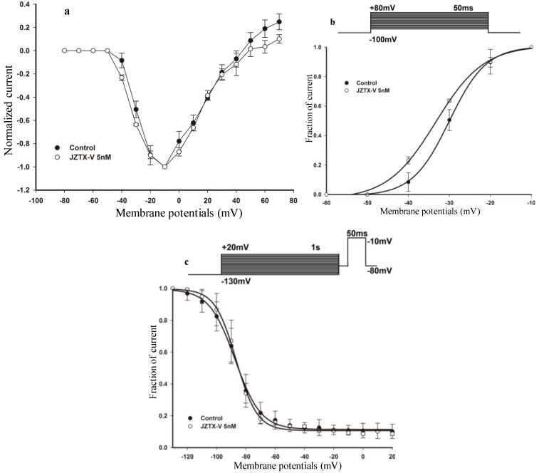
Figure 2.
Effect of JZTX-V on rNaV1.4 channels. (a) Current-voltage (I–V) relationship of sodium currents before (solid circle) and after (open circle) treatment with 5 nM JZTX-V at holding potential for approximately 10 min. All the currents were normalized to the maximum amplitude of peak current; (b) Voltage-dependence of steady-state activation was estimated using a standard protocol. The above diagram shows the protocol for steady-state activation analysis. The currents data used for (a) and (b) were induced by 50-ms depolarizing steps to various potentials from a holding potential of −100 mV. Test potentials ranged from −80 mV to +80 mV in 10-mV increment. HEK 293 cells expressing rNaV1.4 channels were incubated in 5 nM JZTX-V for 10 min at a holding potential of −100 mV to allow toxin binding. JZTX-V (5 nM) shifted the steady-state activation hyperpolarizingly; (c) Voltage-dependence of steady-state inactivation was estimated using a standard double-pulse protocol. The above diagram shows the protocol for steady-state inactivation analysis. The rNaV1.4 currents were induced by a 50-ms depolarizing potential of −10 mV from various prepulse potentials for 1 s which ranged from −130 to +20 mV with a 10 mV increment. JZTX-V (5 nM) had no obvious effect on steady-state activation. Data points (mean ± standard error) from activation and inactivation kinetics were fit with Boltzmann function.

