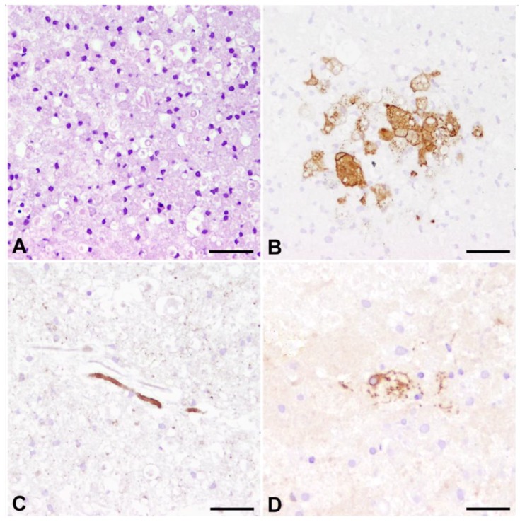
Figure 6.
Organotypic slice cultures of the canine cerebellum, infected with the R252 strain of CDV and cultivated for nine days. (A) Hematoxylin and eosin staining reveals a remarkable response of activated, large phagocytic Gitter cells, presumably originating from resident microglia; (B) Immunohistochemistry for CDV shows that CDV-antigen is predominantly expressed by large, phagocytic cells reminiscent of microglia/macrophages. Whether these cells are infected by replicating virus or if they have phagocytozed viral antigen, remains to be determined in future studies; (C) Highly similar to axonopathy in spontaneous CDV-DL, axonal damage is characterized by enhanced expression of non-phosphorylated neurofilament. Similar axonal expression of n-NF is however also observed in non-infected slices, presumably due to mechanical transection of axons as a result of slice preparation; (D) p75NTR-positive bi- to multipolar cells emerge in response to prolonged culturing, potentially indicating occurrence of pre-myelinating Schwann cells. (B–D) Avidin-Biotin-Peroxidase Complex Method, 3.3’diaminobenzidine tetrahydrochloride. Bars = 50 µm.

