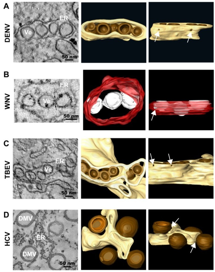
Figure 2.
Representative images of membrane rearrangements induced by different members of the family Flaviviridae. (A) Dengue Virus (DENV); (B) West Nile Virus (WNV); (C) Tick-borne Encephalitis Virus (TBEV); (D) Hepatitis C Virus (HCV). Slices through tomograms of infected cells (on the left) and 3D top and lateral (90° rotation) views of the same tomograms (on the right) are depicted, showing the characteristic virus-induced structures. The replication vesicles (Ve) of DENV, WNV and TBEV (genus Flavivirus) correspond to invaginations of ER membranes that remain connected to the cytosol via 10 nm-pores (highlighted with white arrows in the 3D lateral views), forming vesicle packets (VPs). The replication factory of HCV (genus Hepacivirus) is primarily composed of double membrane vesicles (DMVs) that seem to be formed asER protusions connected to ER membranes via neck-like structures (highlighted with white arrows in the 3D lateral view). The ER is shown in yellow (DENV, TBEV and HCV) or in red (WNV) and the replication organelles in brown (DENV, TBEV and HCV) or in white (WNV). The outer and inner membranes of DMVs are depicted in different shades of brown (outer membrane in dark brown and inner membrane in light brown). Figure 2B is reproduced with permission from [20].

