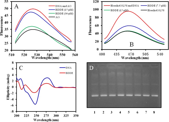
Figure 6.
Interaction mode between BDDE and DNA. (A) BDDE displaces AO from DNA. AO (0.5 μM) was incubated with ctDNA (50 μM) in the absence or presence of BDDE at a concentration of 15, 30 μM for 30 min at 37 °C, respectively. Fluorescence emission spectra (λexc = 502 nm) were determined using a Hitachi F-4500 fluorescence spectrophotometer (Tokyo, Japan); (B) BDDE displaces Hoechst33258 from DNA. Hoechst33258 (1.5 μM) was incubated with ctDNA (50 μM) in the absence or presence of BDDE at a concentration of 7.5, 15.0 μM for 30 min at 37 °C, respectively. Fluorescence emission spectra (λexc = 352 nm) were analyzed using a Hitachi F-4500 fluorescence spectrophotometer (Tokyo, Japan); (C) Intrinsic CD spectra of ctDNA affected by BDDE. CD spectra of ctDNA alone (1.5 mM) or ctDNA treated with BDDE (12.5 μM) were measured with a JASCO 715 spectropolarimeter (Tokyo, Japan); (D) BDDE does not cleave DNA as detected by the agarose gel electrophoresis. Supercoiled plasmid pBR322 DNA was treated without (lane 1) or with 15.5 (lane 2), 31.3 (lane 3), 62.5 (lane 4), 125 (lane 5), 250 (lane 6), 500 (lane 7), and 1000 μM (lane 8) BDDE for 30 min at 37 °C, respectively. The DNA samples were resolved on 1% agarose, stained with ethidium bromide (1 μg/mL) and photographed under UV light.

