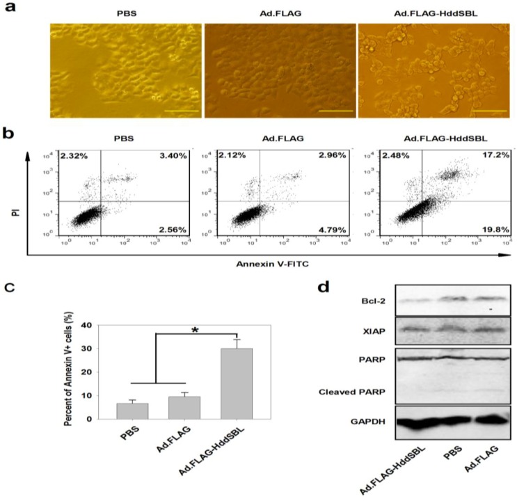
Figure 2.
Ad.FLAG-HddSBL induced apoptosis in Hep3B cells. Hep3B cells treated with Ad.FLAG or Ad.FLAG-HddSBL at 20 MOI as well as PBS control for 48 h. (a) Morphology of apoptosis induced by Ad.FLAG-HddSBL (bars: 200 μm); (b) Cells were stained with Annexin V-FITC and PI followed by analysis under a flow cytometer; (c) The percent of Annexin V-positive cells from three repeats were shown as mean ± SEM (* p < 0.05); (d) Cell lysates were analyzed by Western blot for levels of Bcl-2, XIAP, and PARP. GAPDH served as the loading control.

