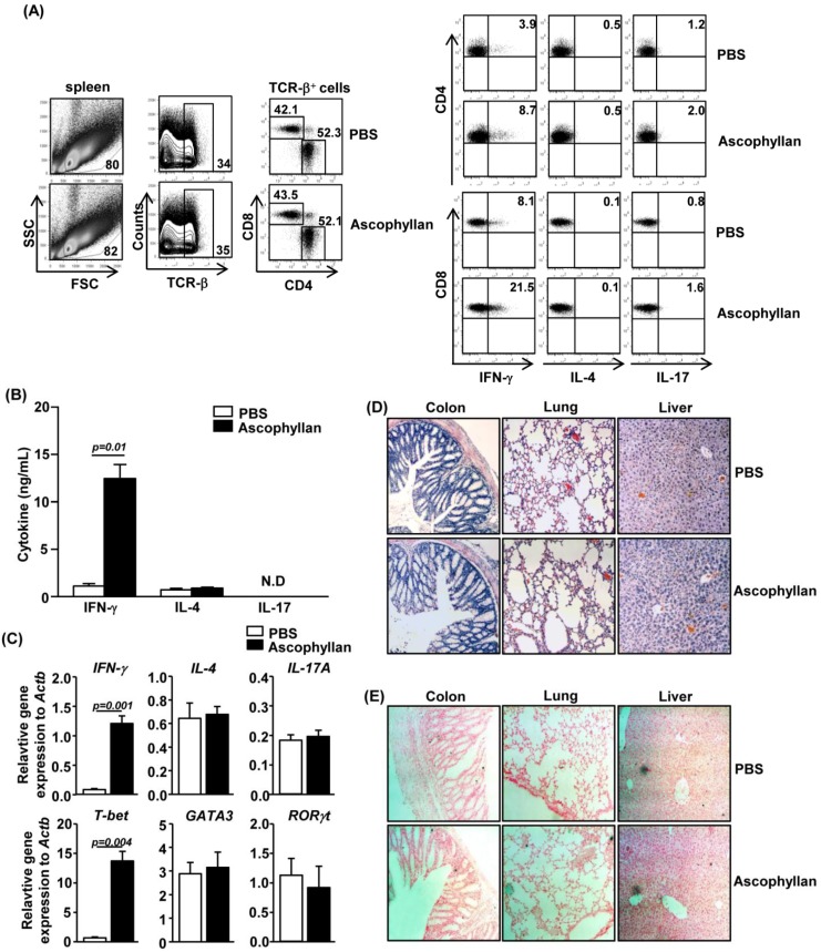
Figure 4.
Ascophyllan promotes IFN-γ production in CD4+ and CD8+ T cells in vivo. C57BL/6 mice were injected i.v. with 50 mg/kg ascophyllan and 3 days later, injected again with the same amount of ascophyllan. Analyses were done 3 days after the second injection. (A) Percentage of IFN-γ-, IL-4- or IL-17-positive cells within CD4+ (right upper panels) and CD8+ T (right lower panels) cells in spleen were assessed by flow cytometric analysis; (B) IFN-γ, IL-4 and IL-17 concentrations in sera were measured by ELISA; (C) Cytokine mRNA levels in spleen were measured 24 h after ascophyllan injection; (D) Hematoxylin and eosin staining of colon, lung and liver sections; (E) In situ detection of cell apoptosis in colon, lung and liver. Arrows indicated the apoptotic cells. All data are representative of six samples from three independent experiments.

