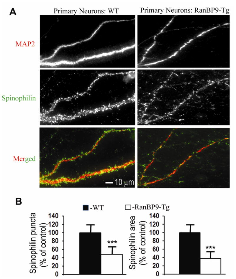
Fig. 4.

Spinophilin-immunoreactive puncta are robustly decreased in primary cortical neurons derived from RanBP9-Tg mice. (A), Primary cortical neurons derived from P0 pups of RanBP9-Tg mice or WT mice were cultured and maintained for 21DIV. At 21DIV, the neurons were co-immunostained for MAP2 to label the dendritic arbor (red) and spinophilin to label spines (green) and the images were acquired in their respective channels. Merged images show spinophilin-immunoreactive puncta on the dendrites. (B), Quantitation of the number of spinophilin-immunoreactive puncta and the spinophilin area by image J showed significantly reduced numbers in the cortical neurons derived from RanBP9-Tg mice compared to WT neurons. Student's t-test revealed significant differences. ***, p<0.001 in the RanBP9-Tg neurons compared to WT neurons. The data are mean ± SEM, n=3 independent experiments with 12 neurons analyzed per experiment.
