Figure 10.
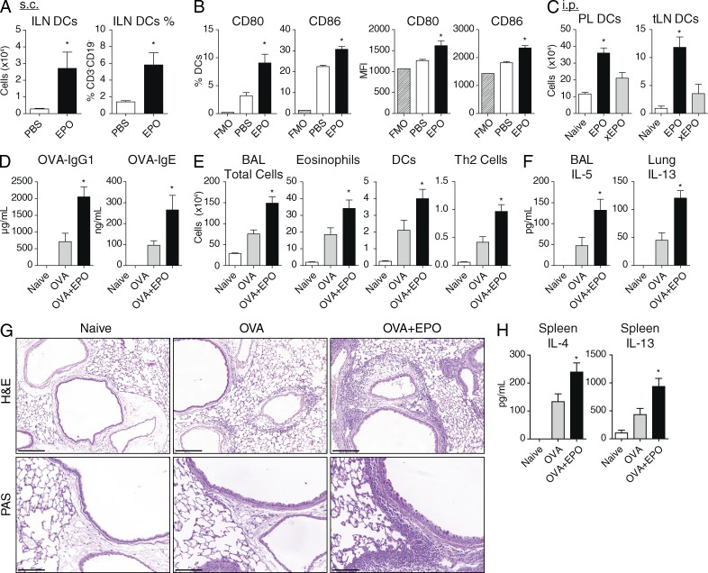
EPO promotes extraintestinal DC and adaptive immune activation in vivo. (A–C) WT mice received PBS, EPO, or heat-inactivated EPO (xEPO) s.c. or i.p. and 24 h later, respective draining inguinal (ILN) or thoracic (tLN) LNs were analyzed. (D–H) WT mice were immunized with OVA alone (OVA) or combination OVA with EPO (OVA+EPO) i.p. on d0 and 14, challenged i.n with OVA on d28-30 and analyzed 24 h later. (D) Serum OVA-specific IgG1 and IgE. (E) BAL inflammatory cells identified by total cell count and flow cytometry. (F) IL-5 in BAL and IL-13 in lung homogenates. (G) H&E staining of lung sections and PAS staining to identify goblet cells. (H) IL-4 and IL-13 cytokine production from OVA-stimulated splenocytes. Mean ± SEM, n = 3–7 from 2 experiments. *, P < 0.05 versus (A and B) PBS, (C) xEPO, or (D–H) OVA alone.

