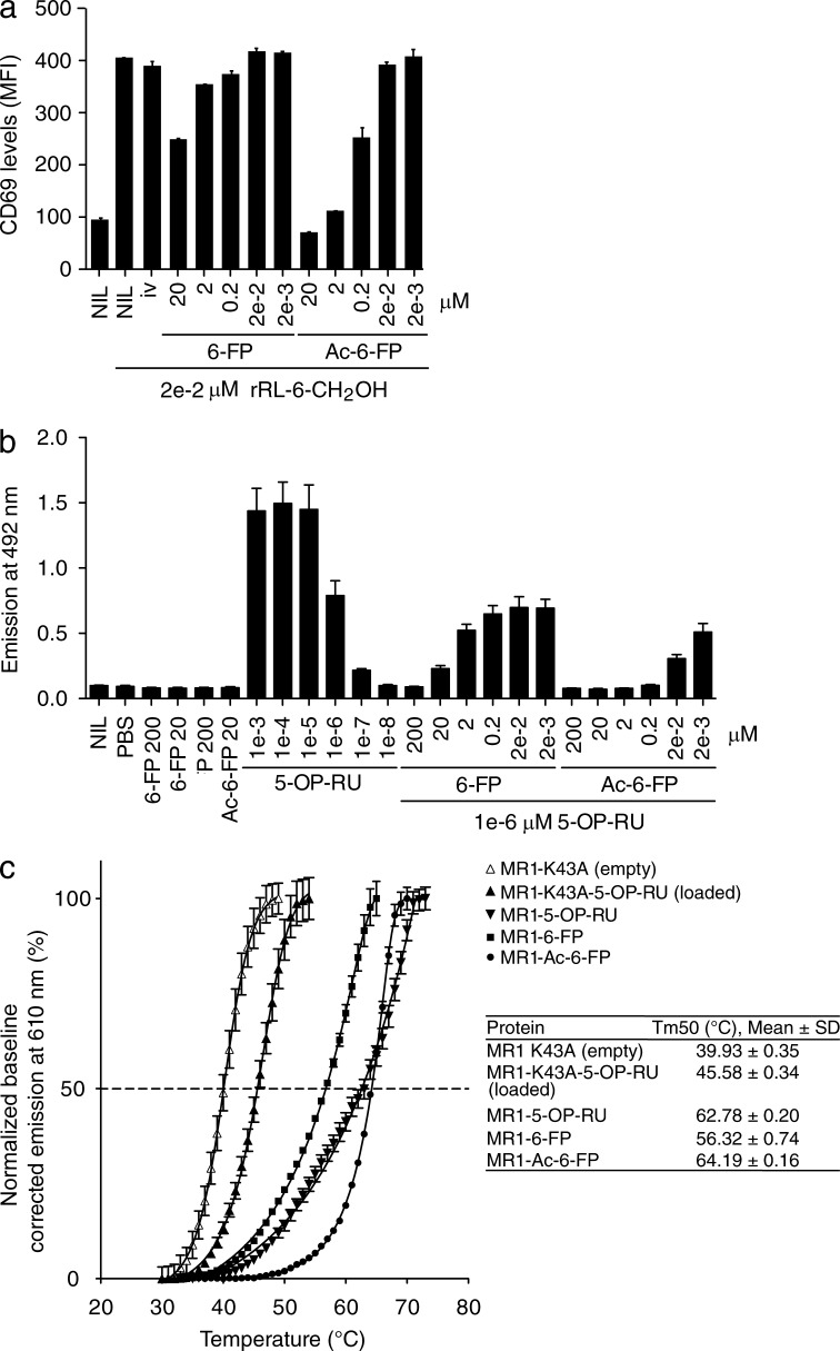
Figure 2.
MAIT cell inhibitory ligands and MR1 stability. (a) Inhibition of Jurkat.MAIT (original WT, clone A-F7) cell activation by 6-FP or Ac-6-FP. 6-FP, Ac-6-FP (or controls – 6,7-diMePterin or no ligand) were added to C1R.MR1 cells at the indicated concentrations for 1 h before addition of Jurkat.MAIT cells and 0.02 µM synthetic rRL-6-CH2OH. Data shows MFI of CD69 expression levels for gated Jurkat.MAIT cells. Mean ± SEM of triplicates. These experiments were performed twice, yielding similar results and a representative experiment is shown. (b) Inhibition of Jurkat76.MAIT (original WT TCR, clone A-F7) cell activation by 6-FP or Ac-6-FP. 6-FP, Ac-6-FP (or controls, PBS or no ligand) were added to C1R.MR1 cells at the indicated concentrations and co-incubated with Jurkat76.MAIT cells and 0.02 µM synthetic rRL-6-CH2OH. Data shows emission at 492 nm correlating with IL-2 production. These experiments were performed three times, showing mean ± SD. (c) Thermostability of soluble MR1-ligand by fluorescence-based thermal shift assay. Shown is baseline-corrected, normalized emission at 610 nm plotted against temperature. Mean ± SEM of triplicate samples, and nonlinear curve-fits. The half maximum melt point (Tm50) is indicated as a dashed line. Displayed is a representative of three independent experiments yielding similar results. The table summarizes the data of three independent experiments, each in triplicate.

