. 2014 Jul 29;3:e02750. doi: 10.7554/eLife.02750

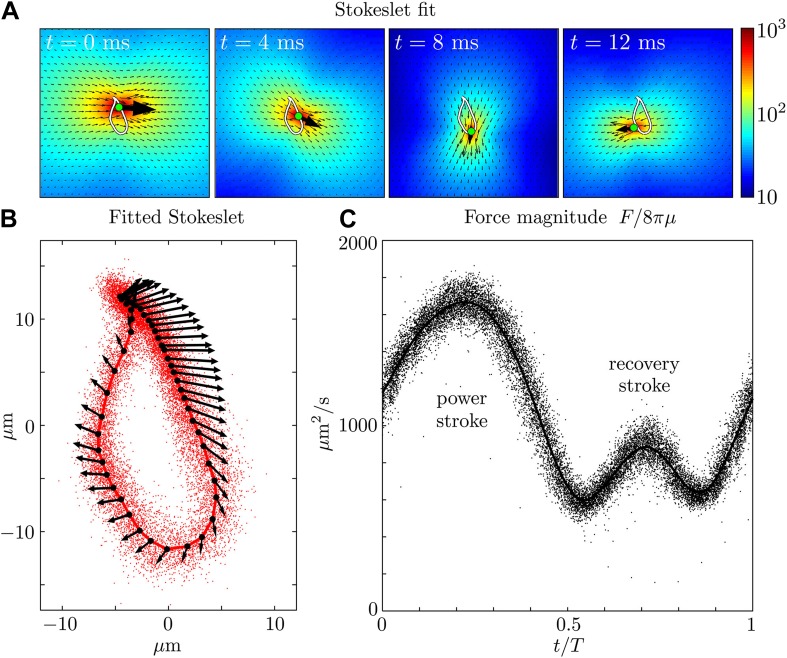
Figure 3. Force amplitude of flagellum.

(A) Fitted instantaneous velocity field at various stages during the first half of one representative flagellar beat. (B) The fitted Stokeslet is shown at evenly-spaced times throughout the average flagellar beat cycle. The red dots indicate the Stokeslet position extracted from every frame. (C) Amplitude of the fitted point force as a function of time throughout the flagellar beat period T.

DOI: http://dx.doi.org/10.7554/eLife.02750.006

Figure 3.

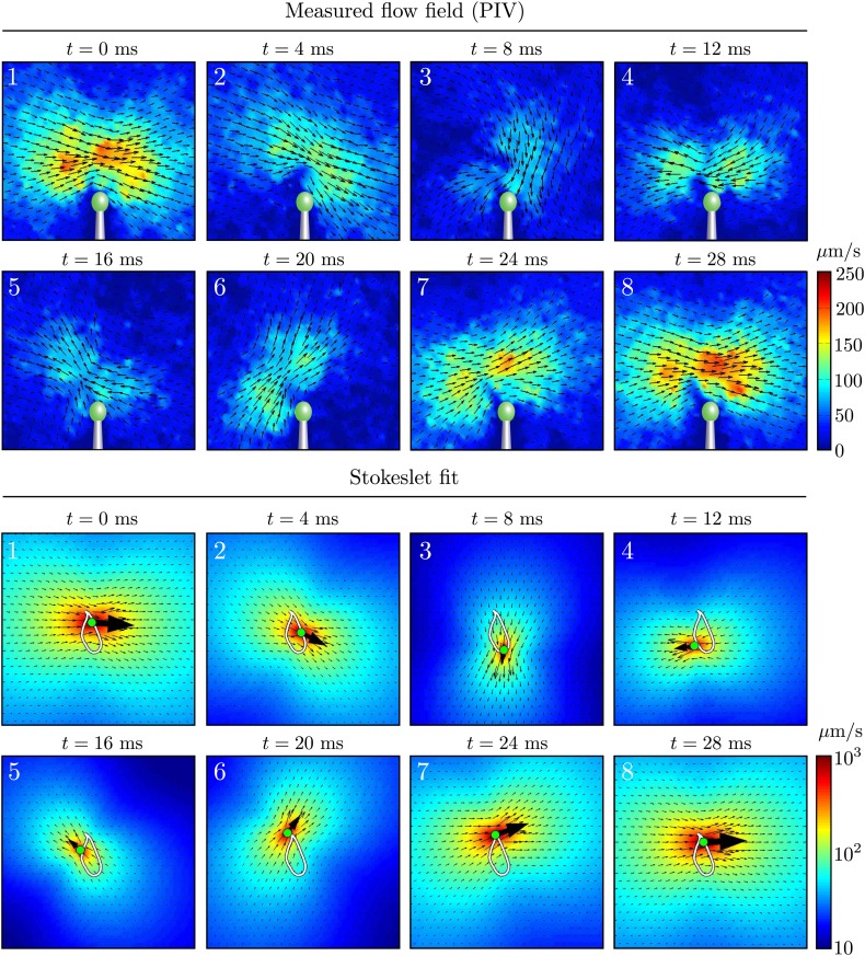
Figure 3—figure supplement 1. Time-dependent flow fields.

Figure 3—figure supplement 1.

Instantaneous fluid velocity corresponding to various stages during one representative flagellar beat. Also shown are the fitted flow fields for each frame, corresponding to the application of a point force on the fluid. Very good qualitative agreement can be seen.