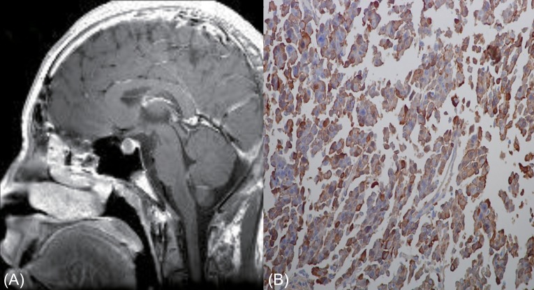
Fig. 1.
Contrast enhanced T1 weighted brain magnetic resonance imaging revealed a 12-mm-sized isointense mass in the pituitary gland (A). A formalin fixed section (B) was immunostained with antibodies against human growth hormone (GH), prolactin, thyroid stimulating hormone beta-subunit, and glycoprotein hormone alpha-subunit. GH-immunopositive cells were common (Immunostain, ×400).

