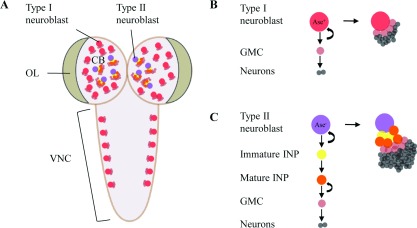
Figure 2. Neuroblast lineages in the Drosophila larval brain.

(A) A dorsal view of Drosophila third instar larval brain which contains three main neurogenic regions: central brain (CB), optic lobe (OL) and ventral nerve cord (VNC). Type I neuroblasts (in red) and type II neuroblasts (in purple) are located at CB. (B) Type I neuroblasts divide asymmetrically to self-renew and produce a GMC (in light red). GMC divides one more time to generate neurons (in grey). (C) Type II neuroblasts divide unequally to generate a self-renewing neuroblast and an immature intermediate neural progenitor (INP, in yellow). After maturation, the INP (in orange) divides asymmetrically to self-renew and generate a GMC.
