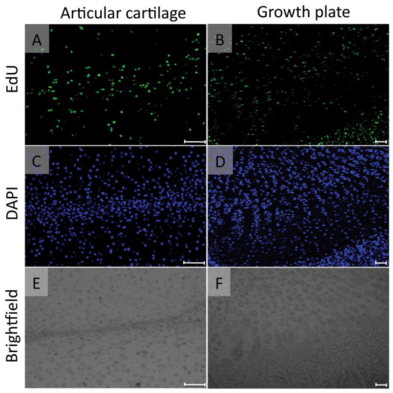
FIGURE 1. Multiple EdU injections labeled most cells in articular cartilage and growth plate.

The mice received 4 daily intraperitoneal injections of EdU (5-ethynyl-2’-deoxyuridine, 5 μg/10 μl/ mouse) from postnatal day 4 and were sacrificed on the next day. The paraffin sections of articular cartilage and growth plate in the medial tibial plateau were subjected to EdU (A and B) and DAPI nuclear (C and D) staining. A large number of the cells in articular cartilage (A-C) and growth plate (B-D) were EdU-labeled. E and F are the brightfield images of A and B, respectively. Scale bars represent 50 μm for A, C and E, 100 μm for B, D and F.
