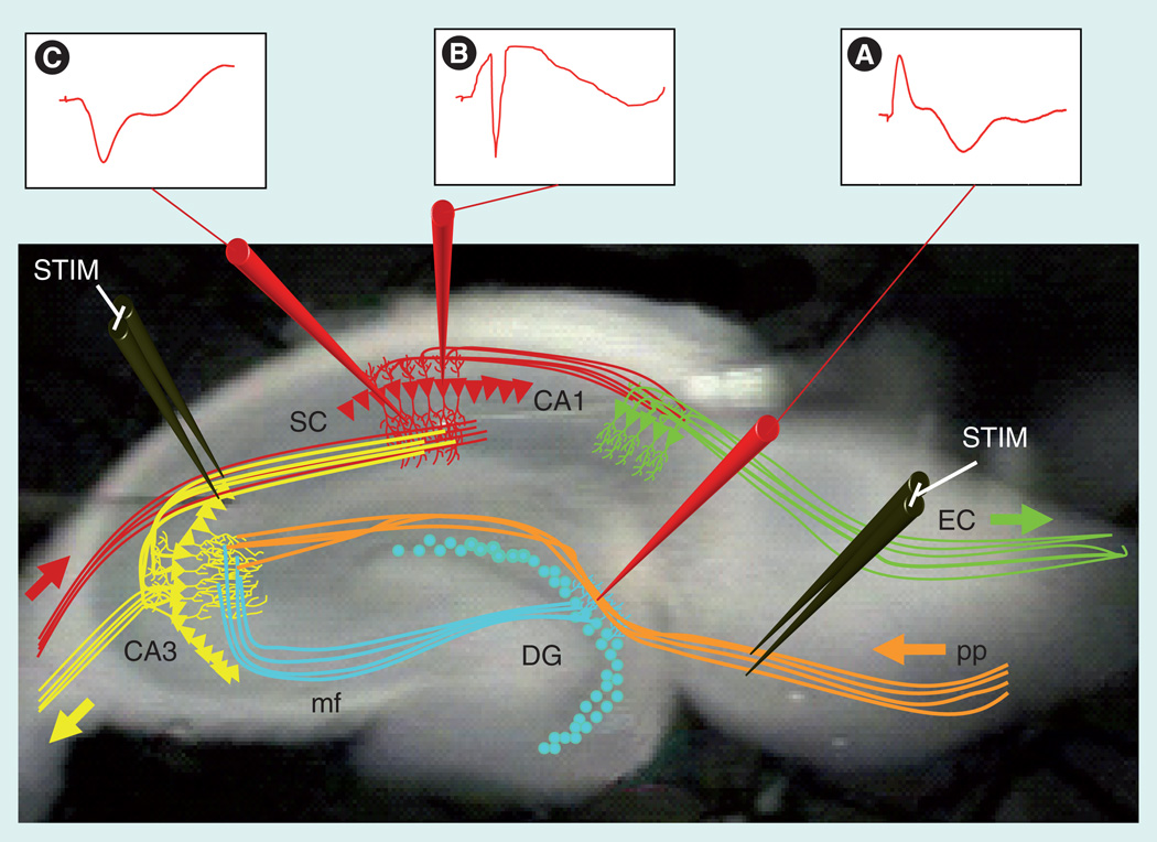
Figure 1. The hippocampal trisynaptic pathway and electrophysiology recording.
The hippocampus consists of a trisynaptic pathway that can be maintained through acute lateral slicing of the mouse brain. Using this technique, the ex vivo slices can be artificially stimulated and recordings can be made across well-known sites of synaptic connections. The hippocampal DG receives major inputs from the EC through activation of the pp. (A) STIM of the pp can be recorded as an excitatory postsynaptic potential (EPSP) in the dendritic field of the DG. The granule cells of the DG synapse onto the dendrites of the pyramidal cells composing area CA3 via the mf. The pyramidal neurons of CA3 project to area CA1 via the SC synapses to the pyramidal neurons of area CA1. Presynaptic STIM of CA3 axons results in EPSPs in area CA1 recorded from either (B) the cell body layer or (C) dendritic fields of CA1. The primary output of the hippocampus is to the subiculum in area CA1, and subsequent signaling exits the hippocampus to the EC.
DG: Dentate gyrus; EC: Entorhinal cortex; mf: Mossy fiber; pp: Perforant path; SC; Schaffer collateral; STIM: Stimulation.

