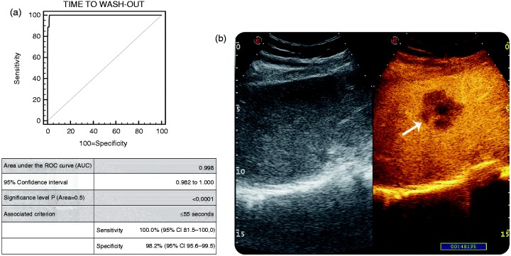
Figure 3.
(a) AUROC for CEUS in detecting non-HCC malignancy within cirrhotic liver in comparison with the gold standard (liver biopsy). (b): B-mode US (on left) and CEUS examination (on right) of FLL in cirrhosis. The early wash-out (i.e. 48 s) was suggestive for non-HCC malignancy (pathologically confirmed to be an ICC).
AUROC: Area under the curve; CEUS: contrast-enhanced ultrasonography; FLL: focal liver lesions; HCC: hepatocellular carcinoma; ICC: intrahepatic colangiocellular carcinoma; US: ultrasonography.

