Fig. 7.
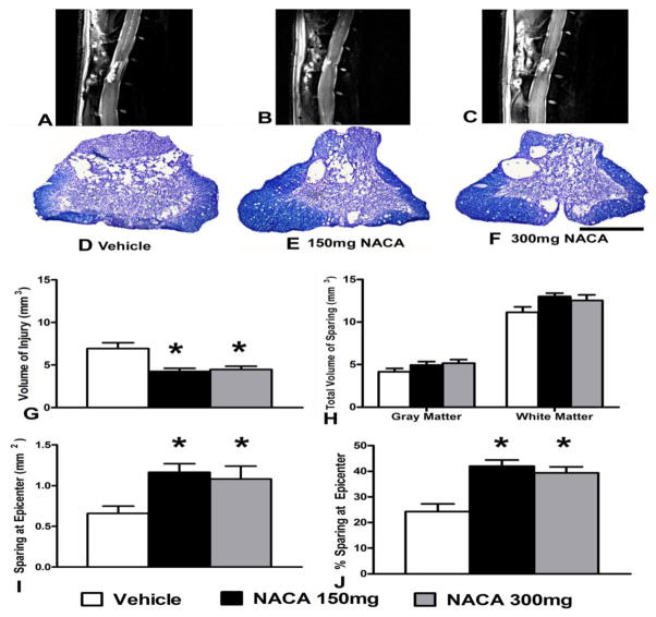
Effects of prolonged continuous NACA treatment on histopathological outcome measures following contusion SCI. (A–C) T2-weighted sagittal MRI images showing reduced lesion volume with NACA treatment compared to Vehicle (white arrows in MRI images indicate injured tissue and yellow arrows pointed at dorsal root ganglion). D–F Photomicrographs of EC- CV-stained injured spinal cord sections from same animals in MRI images, illustrating that the NACA-treated injured groups showed increased sparing of spinal cord tissue at the injury epicenter compared to Vehicle. (G–J) Morphometric quantification of histopathology at 6 weeks post-injury. (G) Both NACA treatment groups showed less volume of injured tissue compared with the Vehicle-treated group. (H) Compared to Vehicle, both gray matter (GM) and white matter (WM) tissues sparing marginally increased with either NACA-treated groups. However, (I) and (J) demonstrate that both NACA treatments significantly increased tissue sparing at the injury epicenter. Bars are group mean ± SEM, n=5–7/group. * p<0.05 compared with vehicle- treated injured group. Scale bar = 1 mm for all histological photos.

