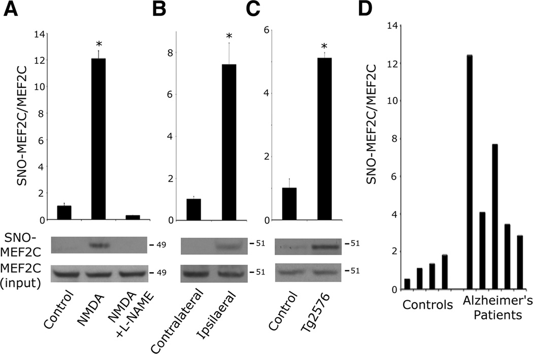
Figure 1. MEF2 is S-Nitrosylated In Vitro and In Vivo during Neurodegeneration.
(A) Excitotoxic NMDA (50 µM) was administered to cortical neurons in the presence or absence of NOS inhibitor l-NAME (500 µM). After 30 min, endogenous SNO-MEF2C was detected by biotin switch. SNO proteins were precipitated from neuronal lysates, and SNO-MEF2C detected with anti-MEF2C-specific antibody. Quantitative densitometry shown above immunoblots. Values are mean + SEM (n = 3 independent experiments, *p < 0.0001 by ANOVA with post-hoc Scheffé's test).
(B) SNO-MEF2C in contralateral or ipsilateral cortex 1 h after MCA occlusion. Values are mean + SEM (n = 3 animals, *p = 0.001 by t test).
(C) SNO-MEF2C in brains from WT littermates and Tg2576 AD mice. Values are mean + SEM (n = 4 animals in each group, *p = 0.001 by t test).
(D) SNO-MEF2C in brain tissues from human control or AD patients by biotin switch.
See also Figure S1.

