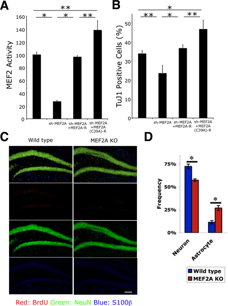
Figure 5. MEF2A is Essential for Adult Hippocampal Neurogenesis In Vitro and In Vivo.
(A) Effect of non-nitrosylatable MEF2 on transcriptional activity during neurogenesis. NSCs were transfected in vitro with MEF2 luciferase reporter, and MEF2A shRNA expression vector, together with expression plasmids encoding shRNA-resistant MEF2A (MEF2A-R) or shRNA and S-nitrosylation—resistant MEF2A [MEF2A(C39A)-R]. MEF2 transcriptional activity was significantly decreased after MEF2A knockdown; MEF2 activity was restored to control levels by MEF2A-R expression and above control levels by MEF2A(C39A)-R. Data are mean + SEM (n = 15 cultures derived from 3 platings, *p < 0.001, **p < 0.01 by ANOVA with post-hoc Scheffé's test).
(B) Effect of non-nitrosylatable MEF2A on neurogenesis. NSCs were transfected as indicated and induced towards neural lineage for 2 d. TuJ1+ cells decreased after MEF2A knockdown. Neurogenesis recovered to control levels with MEF2A-R expression and above control levels with MEF2A(C39A)-R. Data are mean + SEM (n = 15 cultures from 3 platings, *p < 0.001, **p < 0.01 by ANOVA with post-hoc Scheffé's test).
(C and D) Decreased neurogenesis in MEF2A KO mice in vivo. Control and MEF2A KO mice were injected with BrdU and sacrificed 4 week later. Coronal sections of dentate gyrus were immunostained for BrdU (red, newly-proliferating cells), NeuN (green, neuron), and S100β (blue, astrocyte) (C), and stereologically quantified (D). Data are mean + SEM (n = 5 animals for each group, *p < 0.05 by t test). Scale bar, 100 µm.
See also Figure S5.

