
Figure 3. Fluorescence micrographs of Dy547 conjugated miRNA incorporated into gelatin nanofibers.
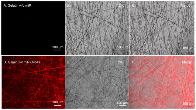
A–C.) Unloaded gelatin nanofibers and D–F.) gelatin nanofibers loaded with fluorescently labeled miRNAs. Differential interference contrast (DIC) image, fluorescent miRNAs (red) (scale 100 μm).