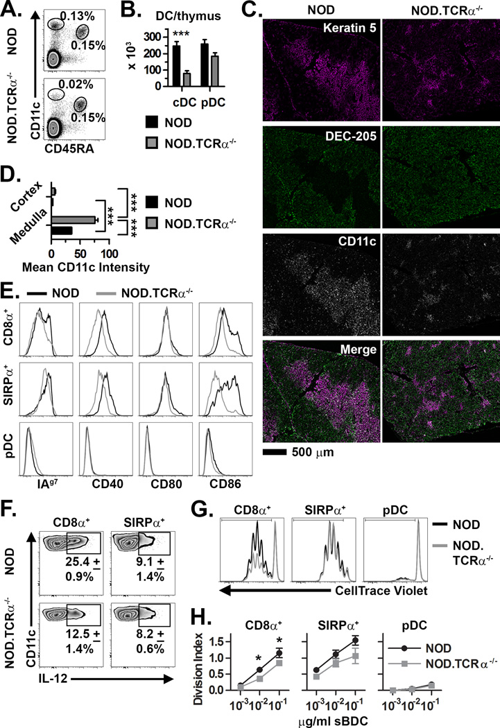
Figure 1. Dysregulation of thymic DC in NOD mice lacking SP.
(A) Frequency and (B) absolute number (±SEM) of thymic cDC and pDC in NOD and NOD.TCRα−/− thymi (n=8 each). (C) Staining of thymic sections for Keratin 5+ mTEC, DEC-205+ cTEC, and CD11c+ DC. (D) Quantification of mean CD11c intensity per unit area (±SEM) in the thymic medulla and cortex (n=12 sections each). (E) Analysis of MHC and costimulatory molecule expression by NOD and NOD.TCRα−/− thymic DC. Data are representative of 4 experiments. (F) Constitutive intracellular IL-12 expression (±SD from 3 experiments) by thymic cDC from NOD and NOD.TCRα−/− mice. (G) DC subsets were FACS-sorted from NOD and NOD.TCRα−/− thymi and BDC2.5 CD4+ T cell proliferation measured. Histograms are gated on live/Thy1.2+/CD4+ cells from representative co-cultures with 10−2 µg/ml sBDC-pulsed DC subsets. (H) Division Index (±SEM) calculated from cells proliferating in Panel G. Data represent 3 pooled experiments. *, P<0.05; ***, P<0.001.

