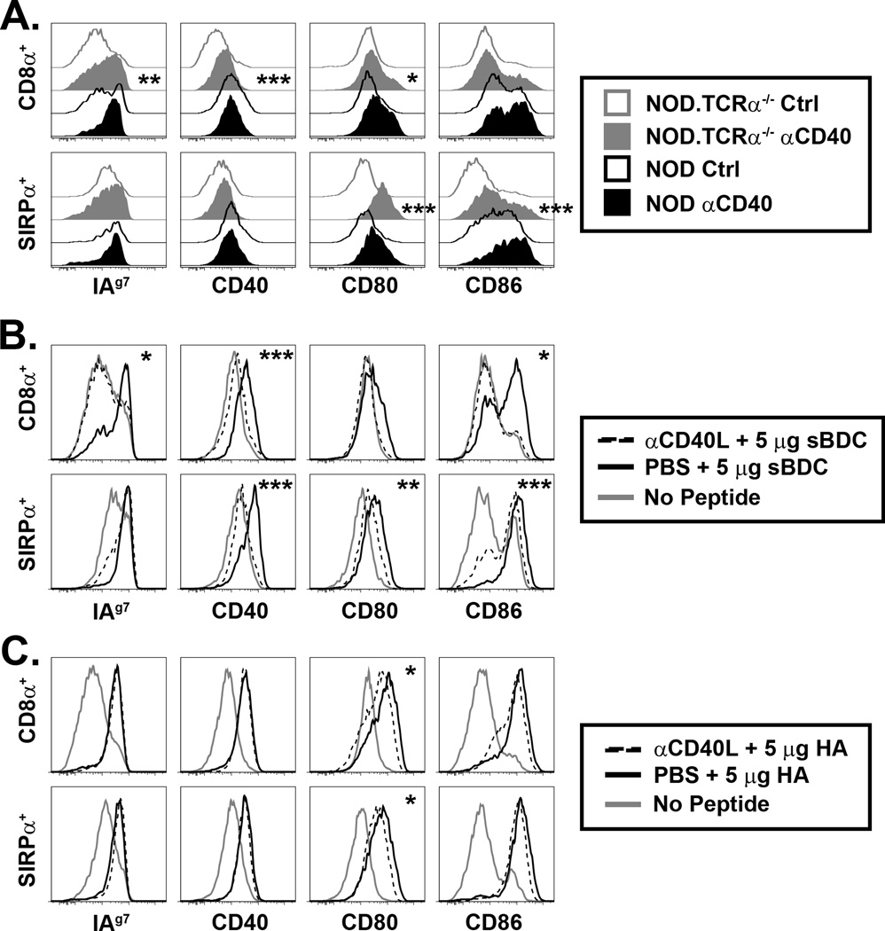
Figure 3. CD40/CD40L partially regulates thymic DC phenotype.
(A) NOD and NOD.TCRα−/− mice were injected i.p. with 200 µg agonist αCD40 or isotype control (Ctrl) mAb, and MHC and costimulatory molecule expression by thymic DC assessed 16–18 h later. Inset asterisks represent analysis of Ctrl vs. αCD40. (B) BDC2.5/TCRα−/− or (C) CL4.scid mice were treated daily i.p. for 3 d with 250 µg blocking αCD40L mAb or PBS then, at the time of the final αCD40L treatment, injected i.v. with 5 µg sBDC (B) or 5 µg HA (C), and thymic DC expression of MHC and costimulatory molecules measured 16–18 h later. Inset asterisks represent analysis of PBS + peptide vs. αCD40L + peptide. Data are representative of 3–5 experiments. *, P<0.05; **, P<0.01; ***, P<0.001.

