Figure 1.

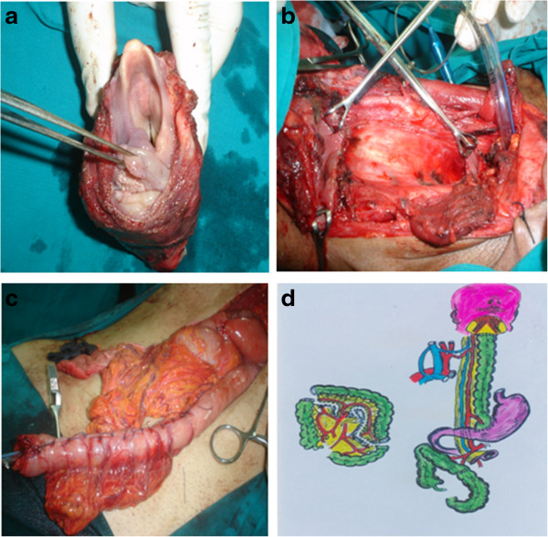
Operative steps and schematic diagram. (a) Specimen after removal. (b) Empty neck after specimen removal. (c) Reversed gastric tube ready for anastomosis in the neck. (d) Schematic view to the augmented colon by pass.

Operative steps and schematic diagram. (a) Specimen after removal. (b) Empty neck after specimen removal. (c) Reversed gastric tube ready for anastomosis in the neck. (d) Schematic view to the augmented colon by pass.