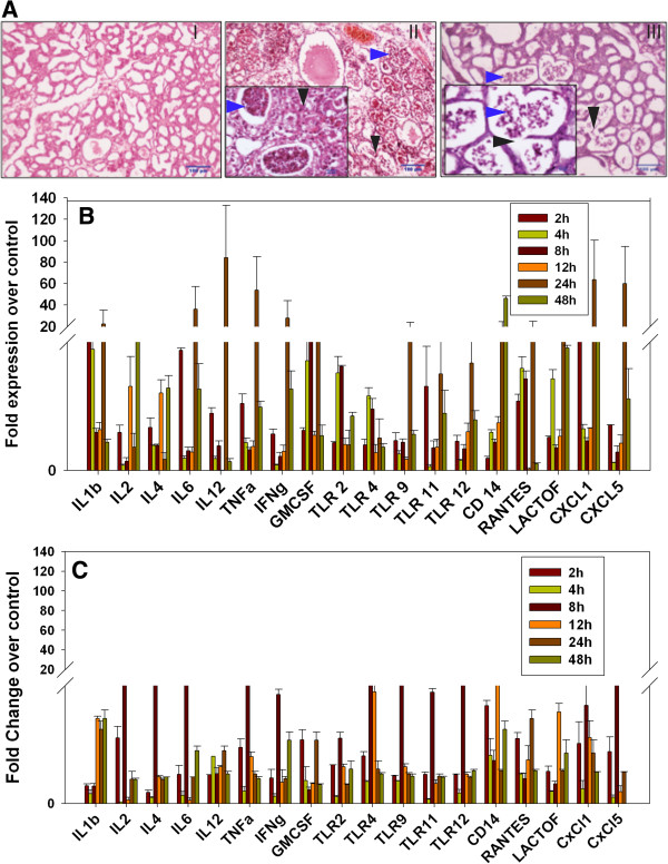
Figure 2.

S. aureus infection induces mastitis in mice mammary tissue. (A) Comparative histopathological analysis of PBS (I) versus S. aureus (II and III) inoculated mice mammary tissue showed clear induction of mastitis after 24 h of infection. SA1 (panel II) inoculated tissue showed more pronounced infiltration of macrophages and neutrophils (blue arrow) in the alveolar lumen, than SA2 (panel III). Black arrow indicates tissue necrosis. (B,C) Temporal expression of various genes in infected tissue against PBS control was analyzed by qRT-PCR. Fold overexpression of the genes in S. aureus infected tissue over the control tissue was calculated and plotted as average of three biological replicates. SA1 (B) infection consistently showed higher inflammatory response compared to SA2 (C) at all time points.
