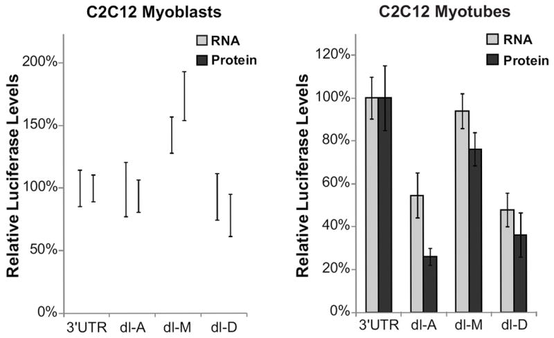
Figure 4. The conserved regions in the DMD 3’UTR increase translation in C2C12 myotubes.

Renilla mRNA and protein levels for C2C12 myoblasts and myotubes transfected with the full-length 3’UTR construct (3’UTR) or deletion constructs (dl-A, dl-M, dl-D) are shown. mRNA levels were measured using qPCR relative to a co-transfected pHRL control. Protein levels were measured using a dual-luciferase assay as previously described. mRNA and protein levels were normalized to the amount of mRNA and protein levels of the full-length 3’UTR. The average of at least three biological transfection replicates is shown for each construct. Error bars equal +/- 1 standard deviation.
