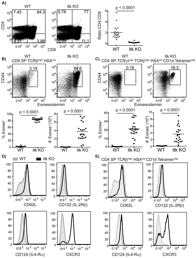
Figure 1. Mature Eomesodermin+ CD8 and CD4 T cells develop in the absence of Itk.
Thymocytes from WT and itk-/- mice were isolated and stained with CD1d-tetramer and antibodies to CD4, CD8, TCRβ, TCRδ, HSA (CD24), CD44, CD62L, CD122 (IL-2Rβ), CXCR3, CD124 (IL-4Rα), and Eomesodermin.
(A) CD4:CD8 ratio of total thymocytes in WT versus itk-/- mice.
(B-C) Eomesodermin versus CD44 staining of CD8SP (B) and CD4SP (C) thymocytes, gated as indicated. The numbers indicate the percentages of CD44high Eomes+ cells in each subset. The graphs below show compilations of all data indicating percentages and absolute numbers of Eomes+ cells in each subset. n = 20-22 mice per group from more than five independent experiments. Statistical analysis was done using a Mann-Whitney test.
(D-E) Histograms show staining of CD62L, CD122, CD124, and CXCR3 on mature CD8SP (D) or mature CD4SP (E) thymocytes. WT thymocytes are shown in gray filled histograms, itk-/- thymocytes are shown in black. (D) WT and itk-/- thymocytes are gated on TCRβ+ CD24low CD8 SP cells. (E) WT thymocytes gated on CD4 SP TCRβ+ CD1d-tetramerneg HSAlow thymocytes, itk-/- thymocytes gated on CD4 SP TCRβ+ CD1d-tetramerneg HSAlow Eomesodermin+ thymocytes. Results are representative of at least three independent experiments.

