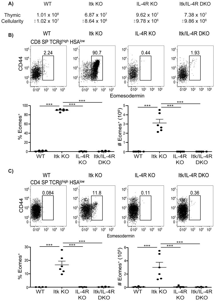
Figure 6. CD4+ and CD8+ Eomes+ T cells are dependent on IL-4.
Thymocytes from WT, itk-/-, il4ra-/-, and itk-/-il4ra-/- mice were stained with antibodies to CD4, CD8, TCRβ, HSA, CD44, and Eomesodermin. Dot-plots show Eomes versus CD44 staining, and graphs show compilations of the percentages and absolute numbers of Eomes+ cells.
(A) Total thymic cellularity. Significant differences were detected between WT and itk-/- (p < 0.0001), WT and itk-/-il4ra-/- (p < 0.0005), itk-/- and il4ra-/-(p < 0.0005), and il4ra-/- and itk-/-il4ra-/- (p < 0.005).
(B) Gated on CD8SP TCRβhigh CD24(HSA)low thymocytes.
(C) Gated on CD4SP TCRβhigh CD24(HSA)low thymocytes.
n = 3-6 mice per group. Results are representative of three independent experiments. Statistical analysis was performed using one-way ANOVA. ***p < 0.0001

