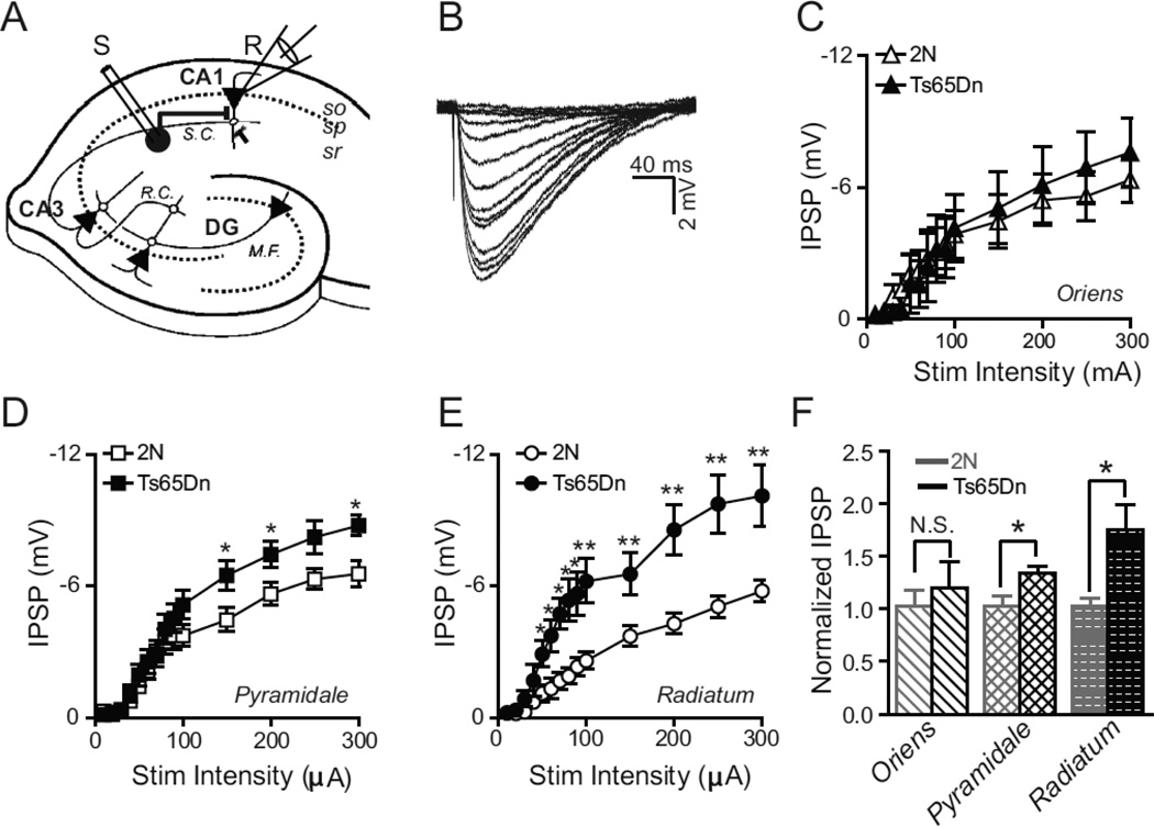
Figure 1. Layer Specific Alteration in Evoked Monosynaptic Inhibition.
(A) Schematic of the hippocampal slice with the whole-cell recording configuration in the CA1 region. Inhibitory interneuron circle in stratum radiatum (sr) of CA1 is stimulated by extracellular stimulating electrode (S) while the inhibitory response is measured from a CA1 pyramidal neuron (triangle) in the stratum pyramidale (sp) by the recording electrode (R). M.F.-mossy fibers, DG-dentate gyrus, R.C.-recurrent collaterals, S.C.-Schaffer collaterals.
(B) Example traces of evoked postsynaptic inhibitory potentials (IPSP) recorded from a CA1 pyramidal neuron in response to various stimulation intensities ranging from 10–300 µA delivered in the stratum radiatum.
(C–E) Plots of evoked IPSP amplitude as a function of stimulus amplitude when the stimulation electrode is placed in the stratum oriens (C), pyramidale (D), and radiatum (E), respectively. There was a significant excess of evoked inhibition in Ts65Dn animals as compared to 2N littermates when the stimulating electrode was placed in s. radiatum (E) and s. pyramidale (D) but not in s. oriens (C). 2N, n=8; Ts65Dn, n=7, where ‘n’ refers to the total number of animals; *P <0.05; **P <0.001, ANOVA). Error bars represent SEM.
(F) Inhibitory synaptic strength in Ts65Dn and 2N animals, in each layer, normalized to the average maximal synaptic strength (at 300 µA stimulation intensity) in 2N animals. * P <0.03; unpaired, two-tailed t-test. Error bars represent SEM.

