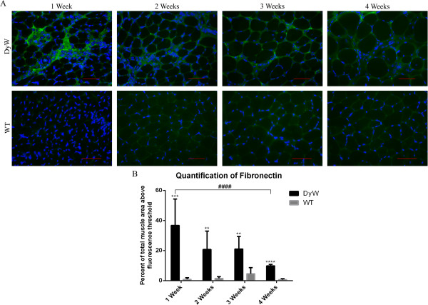
Figure 8.

Qualitative analysis of fibronectin 1–stained frozen tibialis anterior muscle sections from DyW and WT mice at postnatal weeks 1 through 4. (A) Qualitative analysis shows that the highest levels of fibronectin in the extracellular matrix of Lama2DyW (DyW) mice appear to have occurred at postnatal week 1. WT, Wild type. (B) Quantification using fluorescence thresholding confirms the immunohistochemical results and shows that DyW mice have elevated fibronectin expression at each time point (P < 0.01 by t-test (n = 3)). Though this expression decreased with age, fibronectin remained persistently upregulated throughout postnatal development. Bar = 50 μm.
